Diagnostic Equipment
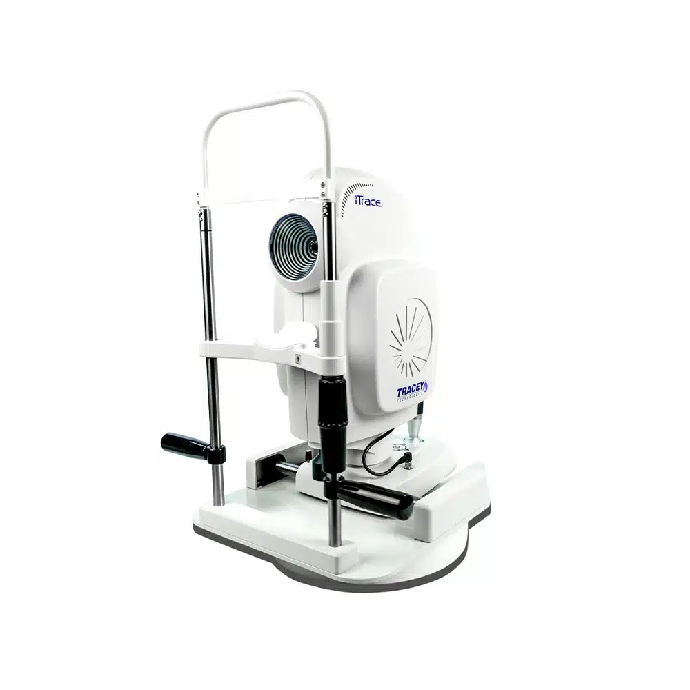
iTrace
The iTrace system is an advanced diagnostic tool that analyzes how light travels through your eye. It helps your doctor evaluate the cornea and internal lens to identify issues affecting visual quality, such as aberrations or early cataracts. This detailed information helps guide personalized treatment and surgical planning.
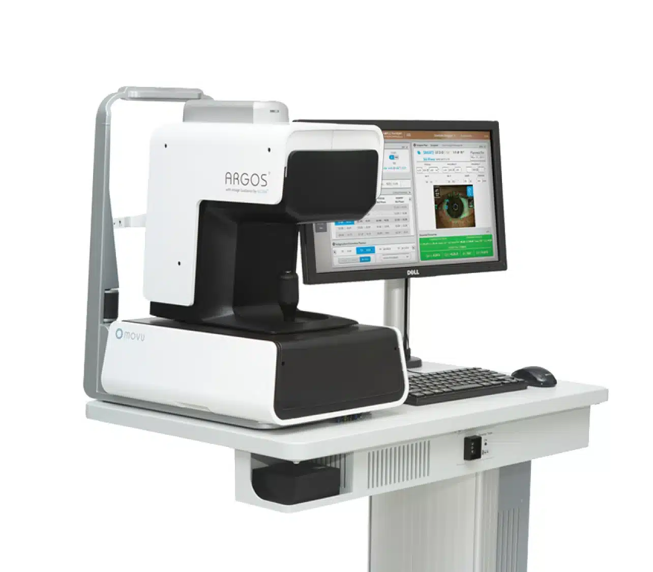
Argos
Argos is a high-precision optical biometer used to measure the eye before procedures such as cataract surgery. It uses advanced imaging to calculate the ideal lens implant for your eye. These precise measurements help improve surgical accuracy and visual outcomes.
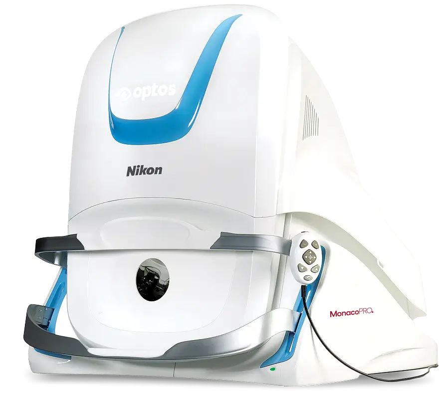
Optos / Optomap
Optos ultra-widefield imaging captures a detailed view of the retina in a single image. This technology allows your doctor to examine more of the retina than traditional methods, helping detect conditions such as retinal tears, diabetic eye disease, and macular degeneration. The scan is quick, comfortable, and often performed without dilating your eyes.
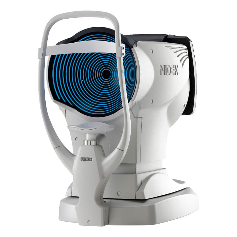
OPD Scan III
The OPD Scan III provides a comprehensive analysis of your vision and the optical system of the eye. It measures refractive errors, corneal shape, and visual distortions to help determine the best treatment options. This technology is particularly helpful when evaluating patients for vision correction procedures.
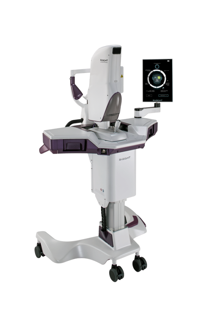
Rx LDD
The Rx LDD device measures tear film stability and ocular surface health. This information helps diagnose dry eye and other surface conditions that can affect comfort and vision quality. Understanding tear film health allows doctors to recommend more targeted treatments.
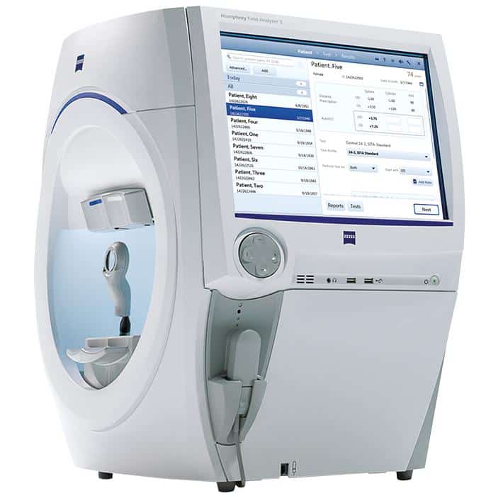
Humphrey Field Analyzer
The Humphrey Field Analyzer 3 is the gold standard for measuring a patient’s visual field. It evaluates how well you see in your peripheral vision, which is essential for detecting and monitoring conditions such as glaucoma and neurological disorders. The test is noninvasive and helps track changes in vision over time.
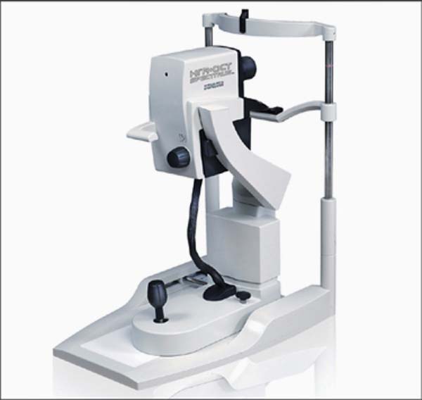
Heidelberg HRA + OCT (Spectralis)
The Heidelberg Spectralis combines optical coherence tomography (OCT) with retinal imaging to create highly detailed cross-section images of the retina. This technology allows doctors to detect and monitor conditions such as macular degeneration, glaucoma, and retinal swelling with exceptional precision. It plays an important role in early diagnosis and ongoing eye health management.